
CLINICAL VALUE
Identifying disease activity through metabolic imaging, beyond structural changes
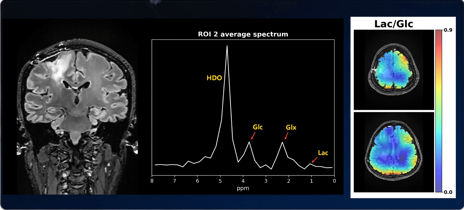
Tumor metabolism is closely linked to the Warburg effect. MR deuterium metabolic imaging sensitively detects metabolites such as lactate, adding a new dimension for early detection, efficacy assessment and mechanistic research.
Core Mechanism
From glucose uptake to energy generation, directly observing the full process of cellular metabolism
After entering cells, deuterium-labeled glucose participates in aerobic glycolysis and downstream mitochondrial oxidative phosphorylation. ²H-MRSI can detect signals such as ²H-Glucose, ²H-Lactate, ²H-Glutamate and HDO, probing the bias of cellular metabolic pathways and changes in flux.
Application Scenarios
Covering early screening of major diseases, efficacy assessment and drug development
Early detection of major diseases
Fast, precise efficacy assessment
Mechanism & target research
Personalized precision care
Technology Evolution
Metabolic imaging becomes a frontier of MR
MR has progressed from structural imaging and functional imaging toward metabolic imaging, with the observation scale moving from tissue morphology to metabolic changes at the cellular and molecular level.
Structural imaging
Observes macroscopic changes in tissue structure or morphology.
Functional imaging
Observes functional changes of tissue at the molecular level.
Metabolic imaging
Observes cellular metabolic changes at the molecular level, becoming a standard direction for next-generation MR products.